Latest

Tuesday, May 19, 2020

What is Diagnostic Radiology and what's it Used For?

No comments:
Current Technology gives doctors many options when it comes to diagnosing a patient’s condition. Some techniques are invasive, others exploratory, and others are minimally or non-invasive. Diagnostic radiology refers to a gaggle of methods that utilize non-invasive techniques to spot and monitor certain diseases.


Diagnostic Imaging
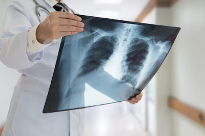
Diagnostic radiology refers to the sector of drugs that uses non-invasive imaging scans to diagnose a patient. The tests and equipment used sometimes involve low doses of radiation to make highly detailed images of a neighborhood.

Examples of diagnostic radiology include:
  • Radiography (X-rays)
  • Ultrasound
  • Computed Tomography (CT) Scans
  • Magnetic Resonance Imaging (MRI) Scans
  • Nuclear Medicine Scans


Diagnostic radiology is often wont to identify a good range of problems. Broken bones, heart conditions, blood clots, and gastrointestinal conditions are just a few of the problems that can be identified by diagnostic imaging.
In addition to identifying problems, doctors can use diagnostic radiology to watch how your body is responding to current treatment. Diagnostic radiology also can screen for diseases like carcinoma and carcinoma.


Technology Used in Radiology
The technology and machinery used in radiology vary from method to method. Some use radiation while others do not.
The most common machines used in radiology are:

X-ray Machine: Uses X-rays, a kind of electromagnetic wave, to supply images of the inside of the body without having to form any incisions.

CT Scanner: Uses X-ray equipment to make a sequence of cross-sectional images of the body. Often used when a doctor needs highly detailed images to review to spot the source of a drag, especially on soft tissue.

MRI Machine: Uses a magnetic flux rather than radiation to supply images of the within of a body. Used for parts of the body that CT scanners cannot produce clear images of, such as bones.
Some of the diagnostic tests may require compounds to be ingested or chemicals to be injected for a transparent view of your blood veins. Other tests may require anesthesia and scope so as for a doctor to obviously determine the matter.

Star Radiology
Star Radiology uses imaging technology like CT scans, MRI, and Ultrasound to assist guide medical procedures. This technology eliminates the need for surgery and scopes to diagnose and treat certain conditions. Instead, patients are often awake during the procedure or under very mild sedation.

Common uses for Star Radiology include:
  • Treating cancers
  • Treating blockages in arteries or veins
  • Treating back pain
  • Treating liver and kidney problems

Star radiology is a pioneer in teleradiology services in India.

Saturday, May 9, 2020

Difference Between X-rays, CT scans and MRI

No comments:
In the Current Era, doctors mainly use diagnostic imaging techniques to narrow down possible causes of pain or illness for a more accurate diagnosis. There are different types of diagnostic imaging, including X-rays, CT scans, and MRI. the sort of imaging used depends on the part of the body the doctor wants to ascertain on a picture, also because the sort of imaging that's readily available to the patient.

So what's the main difference between X-rays, CT scans and MRI?


X-Rays
X-rays are the foremost common and widely available diagnostic imaging technique. albeit a patient may have a more sophisticated test, they're going to often receive an X-ray first.

X-rays use radiation to supply images of the body. When the rays undergo the body, dense objects—such as bones—appear white on the film. X-rays are typically wont to view and diagnose bone disease, degeneration, fractures, dislocations, infections and tumors.

Though often wont to examine skeletal structures, an X-ray also can be wont to check out other internal structures, like organs. during this case, the patient may given barium sulphate or dye to form the organs stand out more clearly within the X-ray image.

During an X-ray, the a part of the body that's being checked out are going to be placed between an X-ray machine and film . The machine then sends electromagnetic waves (radiation) through the body, reflecting the patient’s internal structures on the exposed film.

Although the quantity of radiation used for an X-ray isn't considered dangerous, doctors will take certain precautions if the patient is pregnant.


CT Scans
A computerized tomography scan, or CT scan, is analogous to an MRI therein it produces detailed, high-quality images of the body. The CT scan may be a more sophisticated and powerful X-ray that takes a 360-degree image of internal organs, the spine and vertebrae. Contrast dyes are often injected into the blood to form structures within the body more visible on the CT scan.

A CT scan produces detailed images of organs, bones, soft tissue and blood vessels and may be wont to more easily diagnose cancer, heart condition , appendicitis, musculoskeletal disorders, trauma and infectious diseases.
A CT scanner seems like an outsized box with a tunnel within the center. The patient lies on a table that slides in and out of the tunnel, while the scanner rotates round the patient, producing cross-section images of the body. The technologist performing the scan sits during a separate room with computers on which the pictures are displayed. The technologist can speak with the patient using speakers and microphones.

A CT scan is costlier than an X-ray and isn't always available at small or rural hospitals.


MRI
Magnetic resonance imaging, or MRI, uses a strong magnet and radio waves to make detailed, high-resolution cross-section images of bones and soft structures inside the body. MRI doesn't include radiation, as X-rays and CT scans do, and is usually wont to diagnose bone and joint problems also as torn ligaments and cartilage and herniated discs.
During an MRI scan, the patient lies still on a table that slides into the tube-shaped MRI scanner. The machine then creates a magnetic flux round the patient and pulses radio waves into the world of the body being pictured. The radio waves cause the tissues within the body to resonate. These vibrations are translated into detailed 2D images captured by a special computer virus .
Like an X-ray or CT scan, the MRI is painless, but the machine does produce a bang . Because the MRI uses large magnets, make certain to notify your doctor if you've got metal clips, implants or other metal objects within the body.
MRI scans aren't available in the least hospitals. If your doctor has ordered an MRI, you'll got to attend a delegated imaging center for your scan.

Thursday, May 7, 2020

X-Ray Service Provider

No comments:
X Ray is a form of high energy electromagnetic radiation. The wave length of the electromagnetic radiation determines its energy and properties ranging from radiowaves (longest wave lengths) to microwave, millimeter or terahertz radiation to infrared, visible light to ultraviolet, X Rays and gamma rays.
X Rays are produced with beams of electrons in a vacuum tube strike a metal and decelerate emitting energy as X Rays.
Due to their high energy they can pass through the soft tissues of our body easily allowing us to see the skeleton inside (conventional X Ray).
DEXA scans that measure the Calcium inside our bones and CT scans are advanced applications of X Rays.


Copyright ©2016 Star Radiology • All Rights Reserved.
Designed By Onlystudybyte